In vitro Traumatic Brain Injury (TBI) model based on Human 3D neural tissues
Ingénierie et Architecture
METHODS
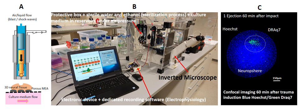
The in vitro TBI device is inducing injury by a fine tunable (speed and force) expulsion of culture medium onto the minibrain by using a microvalve (SMLD 300G, Fritz Gyger AG).
The brain spheroids are generated by non-directed differentiation of neural stem cells derived from human iPS cells (NSChiPS)3. Platform&Trauma: The in vitro TBI device is inducing injury by a finely tunable (speed and force) expulsion of culture medium onto the minibrain (Fig. 1A). The validation of the in vitro TBI platform is done by using complementary methods: electrical monitoring, imaging (confocal microscopy and optical projection tomography), and circulating biomarkers analysis by multiplexing assays4.
RESULTS
The functional electrophysiological monitoring shows an abolition of spike activities just after the injury, followed by induction of burst activities (Fig. 1B). We observe an increase in astrogliosis biomarkers (GFAP) released in the culture medium after the impact (Fig 1C). The injured area is then 3D spatially mapped within the injured minibrain by Optical Projection Tomography (OPT) (Fig. 1D) and confocal microscopy (Fig. 1E) after staining with cell-permeant and non-permeant nuclear dye.
DISCUSSION & CONCLUSIONS
We demonstrate that our innovative combination of human 3D minibrains and in vitro device can mimic brain injury. With these tools, we can evaluate the metabolic changes occurring after impacts, such as electrophysiological dysregulation, tissue loss, and biomarker release. As a perspective, we hope to be able to track mTBI over time and gain a better understanding of the kinetic of biomarker release and cells recovery.
REFERENCES
1 A.R. Mayer et al (2017) The spectrum of mild traumatic brain injury: A review. Neurology 89, 623–632. 2 N. Osier et al (2016) The Controlled Cortical Impact Model of Experimental Brain Trauma: Overview, Research Applications, and Protocol in Injury Models of the Central Nervous System vol. 1462 177–192. 3 S. Govindan, L. et al (2021) Mass Generation, Neuron Labeling, and 3D Imaging of Minibrains. Front. Bioeng. Biotechnol. 8.4 M. Jović, et al (2022) Towards a Point-of-Care (POC) Diagnostic Platform for the Multiplex Electrochemiluminescent (ECL) Sensing of Mild Traumatic Brain Injury (mTBI) Biomarkers. 21