2.0 MR-Eye - A-EOG Eyes Position Detection System to support Sharp MRI of the Eyes
Ingénierie et Architecture
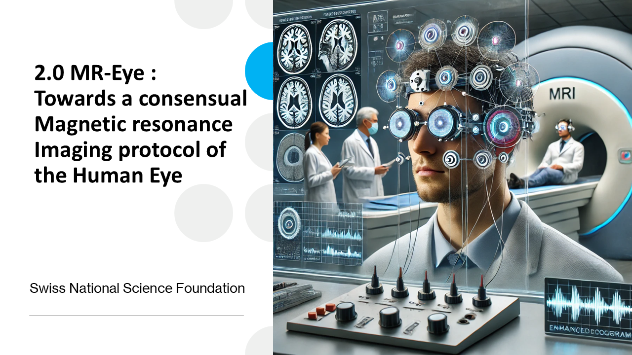
The global project “2.0 MR-Eye: Towards a consensual Magnetic Resonance Imaging Protocol of the Human Eye”, funded by Swiss National Science Foundation, aims at improving human eyes magnetic resonance imaging (MRI) resolution for diagnosis purpose and medical science research, despite eye movements causing image blurring (MRI images require multiple image acquisitions/repetitions averaging). 2.0 MR-Eye project aims at reconstructing sharp eyes images with the support of different eye position tracking technologies allowing spatial resynchronization of eyes MRI scan repetitions.
A reference system used to track and record the eye position in the MRI is the camera-based EyeLink® 1000 Plus system from SR Research Ltd. Which requires quite some installation and calibration time on the patient and whose operation is limited to the tracking of a single eye position (whereas MRI simultaneously scans the two eyes).
The advanced electrooculography (A-EOG) system proposed here is based on the monitoring of biopotentials (very low amplitude electrical signals) surrounding the eyes and varying whenever the eyes are moving. Electrodes are located around the patient’s eyes to monitor these biopotentials. The A-EOG features more electrodes vs a standard EOG system and should allow for more accurate eyes positions detection using smart electrode signals combinations. This system design allows simultaneous monitoring of the two eyes’ positions and should allow for quick installation and calibration time.
Challenges linked to the proposed A-EOG relate to system performance, safety and electromagnetic compatibility within 1.5T, 3T and 7T MRI environments characterized by 1.5T up to 7T static + variable gradient magnetic fields + high magnitude magnetic excitation pulses from 64MHz to 300MHz. To avoid any ballistic effect inside the MRI, the A-EOG structure design is free from ferromagnetic materials. The A-EOG system structure, wiring and location inside the MRI environment is designed to avoid risks of burns to the patient, and to avoid induced noisy voltages impacting the eye-tracking performance. Similarly, the A-EOG is designed to avoid causing artifacts on MRI images by avoiding distorting the MRI magnetic field and by reducing electromagnetic noise emissions. The designed system is thus split into an electrically floating and shielded in-situ acquisition electronic subsystem running on batteries and free from ferromagnetic materials, located close to the patient head, and a remote data recording subsystem located outside the MRI environment (in the control room), both being linked through an optical fiber link.
A-EOG specific challenges are:
- corneo-retinal potential varies with luminosity and across patients
- 0.5° eye angular resolution corresponds to differential signals of ~ 5-10µV magnitude
- Probes contact impedance variation with micro-movements may add noise
- Required system bandwidth is from DC to 1kHz for absolute eye position detection
Limited space is available around the patient’s head